Diagnosed Conditions - Complaints- Spinal
Complaints- Spinal
Back Pain
Back pain is extremely common with around 8 out of 10 people in the UK experiencing some form of back pain in their lives. It is also one of the most common reasons for people being off work and for going to see their doctor. It can impact upon all aspects of your life; ability to work, exercise, enjoy your hobbies, sleep and can negatively impact upon your mood. Back pain can arise from a specific incident or trauma or can occur over time due to repetitive stresses or postural issues such as prolonged sitting. It affects everyone differently and can include symptoms such as a sharp pain or general ache, stiffness and restricted movement. Back pain can also involve the neural system and results in shooting or radiating leg pain that is commonly known as sciatica.
It is important to act quickly to treat back pain as it arises to prevent it from becoming chronic or from reoccurring.
The most common form of back pain is non-specific lower back pain and is mechanical in nature. Pain arises in the lower back and is most commonly as a result of a dysfunction or imbalance in the muscles that are responsible for supporting and moving the spinal column. This can result in abnormal movement patterns that can perpetuate the negative cycle of pain and dysfunction.
Back pain can also be secondary to common spinal conditions that your doctor may have mentioned to you if you have had any scans or investigations. These may include;
Disc Herniation
Disc herniation, prolapsed disc or disc protrusion are all names for the same condition. The intervertebral discs sit between the hard bone of the vertebral bodies that make up the spinal column. They act as shock absorbers and allow smooth fluid movement as we bend and twist. A herniated disc is a failure of the core of the disc called the annulus and during compression, the disc can bulge outward.
Cause can be from a sudden forced flexion or torsion but more often is as a result of repetitive movement or prolonged postures in to spinal flexion.
A herniated disc can irritate the nerves and surrounding structures which results in pain. The protrusion can occur to varying degrees which will impact upon the severity of symptoms. These can include;
- pain related to the spinal segment involved-often exacerbated by standing, sitting or walking
- Sciatica
- Pins and needles
- weakness in arm or leg
You may know you have a disc herniation from the results of a scan such as an MRI. If not explained well to you this can often be extremely worrying and can result in fear of moving which may make your pain worse. However research has shown that results from investigations such as an MRI do not always correlate to patient symptoms. In fact disc herniation can often occur with no symptoms at all with a large proportion recovering spontaneously! Your Physiotherapist can help guide you through the management of this condition, optimize healing and recovery and provide you with strategies to manage and prevent a reoccurrence.
Facet Joint Syndrome
Facet joints can become stiff which results in abnormal movement and pain. This stiffness can be as a result of repetitive or excessive rotation, flexion or extension of the spine resulting in degenerative changes and arthritis.
It may also occur as a result of an uncontrolled motion such as a simple twist or awkward movement which results in facet joint motion going beyond your muscle control. This can be termed as a locked facet joint and can occur repeatedly especially if your local supporting muscles are weak.
Facet joints can also experience excessive mobility or hyper-mobility due to fracture or dislocation, however this is uncommon.
Symptoms:
In the acute stage you may experience local muscle spasm that occurs in an attempt to protect the injured facet joint. This can often prevent you from moving your neck or back.
Cervical facet syndrome: neck pain that limits extension and/or rotational movement and is usually experienced on one side. This pain may radiate down in to the shoulder or upper back.
Lumbar facet syndrome: pain in the lower back that is exacerbated by extension such as standing or getting up from a chair. Referred pain can extend in to the lower leg.
Spondylolysis or Spondlylolisthesis
This condition is considered to be a stress fracture that results from repetitive mechanical stress of the part of the spinal vertebrae called the pars interarticularis. This structure is most susceptible to extension and rotational movement with damage developing over time rather than as a result of a single traumatic event. There is increased prevalence seen in the young athletic population.Clinical presentation is based on the region of the spine affected but can include;
- Gradual onset of back pain, worse after exercise or athletic activity
- Pain may radiate in to buttocks
- Pain may be aggravated by extension and/or rotation of the spine
- Relief from rest
Radiculopathy
Radiculopathy can occur in any part of the spine but is most commonly seen in the lower back. It can arise from multiple pathologies to the spinal vertebrae that cause compression of the nerves that exit the spine. This can result in symptoms such as radiating pain, tingling, pins and needles, shooting pain and occasionally weakness. The most common cause of radiculopathy is as a consequence of a herniated disc (discussed above). However stenosis of the vertebra (discussed below) and also tumours are possible causes.
A common well known radiculopathy is called Sciatica. The sciatic nerve (nerve root L4, L5 S1, S2, S3) can become compressed. Pain radiating down to your leg is worsened with movements such as coughing or sneezing (which increase intra-abdominal pressure) or sitting, bending and prolonged standing. Relief is usually achieved through lying down.
Spinal Stenosis
Each single vertebra in your spine is formed in such a way to create a channel that runs through the centre. Through this channel or ‘spinal canal’ runs the delicate spinal cord which is protected by this bony formation. Between two vertebra the spinal cord branches on either side and exits the spine as nerve roots to supply muscle, skin and organs. Spinal stenosis is a condition whereby the spinal canal narrows and the nerve roots and spinal cord can become compressed. Stenosis can occur as a result of many different pathologies such as disc herniation, spondylolisthesis, trauma, rheumatoid arthritis but is usually as a result of chronic degeneration.
Spinal stenosis can occur in any portion of your spine; cervical, thoracic or lumbar. It can occur without any symptoms but with the ageing process, symptoms may develop.
Typical symptoms include;
- Cervical stenosis: Pain in the neck that increases when you flex your neck.
- Burning, tingling, pins and needles and/or numbness in neck, shoulder, arms and hands.
- Clumsiness with fine motor skills or general feeling of weakness.
- Lumbar stenosis: Pain in the lower back that may extend in to buttocks and/or legs.
- Pain may increase during prolonged standing, extending your back in activities such as walking downhill.
- Relief is often found from flexing your lower back.
- You may experience burning, tingling, pins and needles and/or numbness in the lower back, buttocks, and legs.
- Lower back pain, which may be exacerbated with forward bending activities
- Local tenderness over the SIJ
- Pain may extend in to the buttock or thigh
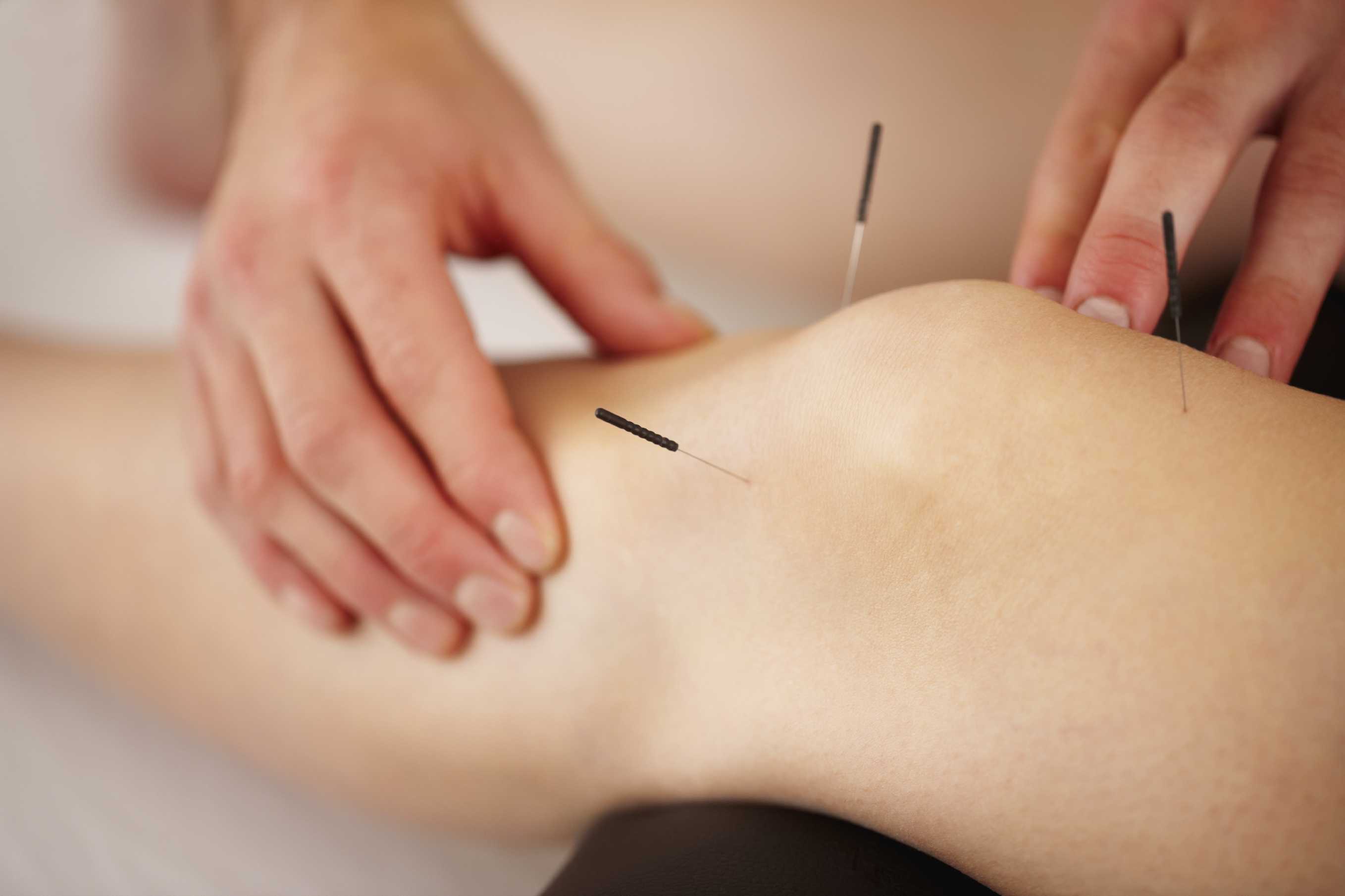
- Specialised hands on Soft-Tissue Techniques, Acupuncture and Stretches to release tension and ease pain,
- Use of Sport Taping to facilitate and help you to achieve correct postures and movement patterns,
- Joint Mobilisations and positions of ease to restore normal pain free movement,
- Techniques to release pressure on affected nerves allowing them to stretch and move normally,
- The use of Electrotherapy equipment such as Class 3b Laser (or low level light therapy) to facilitate healing and restoration of normal tissue. H-wave which uses muscle stimulation to normalise muscle activation.
- Prescribed Exercises to increase your movement and strengthening of the muscles that support your back to prevent back problems reoccurring in the future.
In some instances, such as persisting symptoms or progressive neurological deficits, surgical interventions can be performed to decompress the nervous structures. However non-operative treatment such as physiotherapy should always be explored first.
Sacro-Iliac Joint Dysfunction
The sacro-iliac joints (SIJ) are located between the sacrum and the two pelvic bones. These joints are supported by strong ligaments and muscular attachments. The primary role of this joint is to transmit forces between the lower limbs and the body and to act as a shock absorber for the spine. This joint can be a significant source of pain in those that suffer from lower back pain and can often be overlooked. Dysfunction can occur as a result of traumatic injuries such as a heavy fall landing on the buttocks or seat bone or cumulative injury from repetitive activities such as running. Excessive force to this joint can result in disruption of the supporting ligaments and consequently excessive movement at a joint that doesn’t really move more than a few degrees!
Pregnant women may experience SIJ pain as the hormone ‘relaxin’ released during pregnancy can impact on SIJ ligaments allowing hyper-mobility and pain.
Symptoms include;
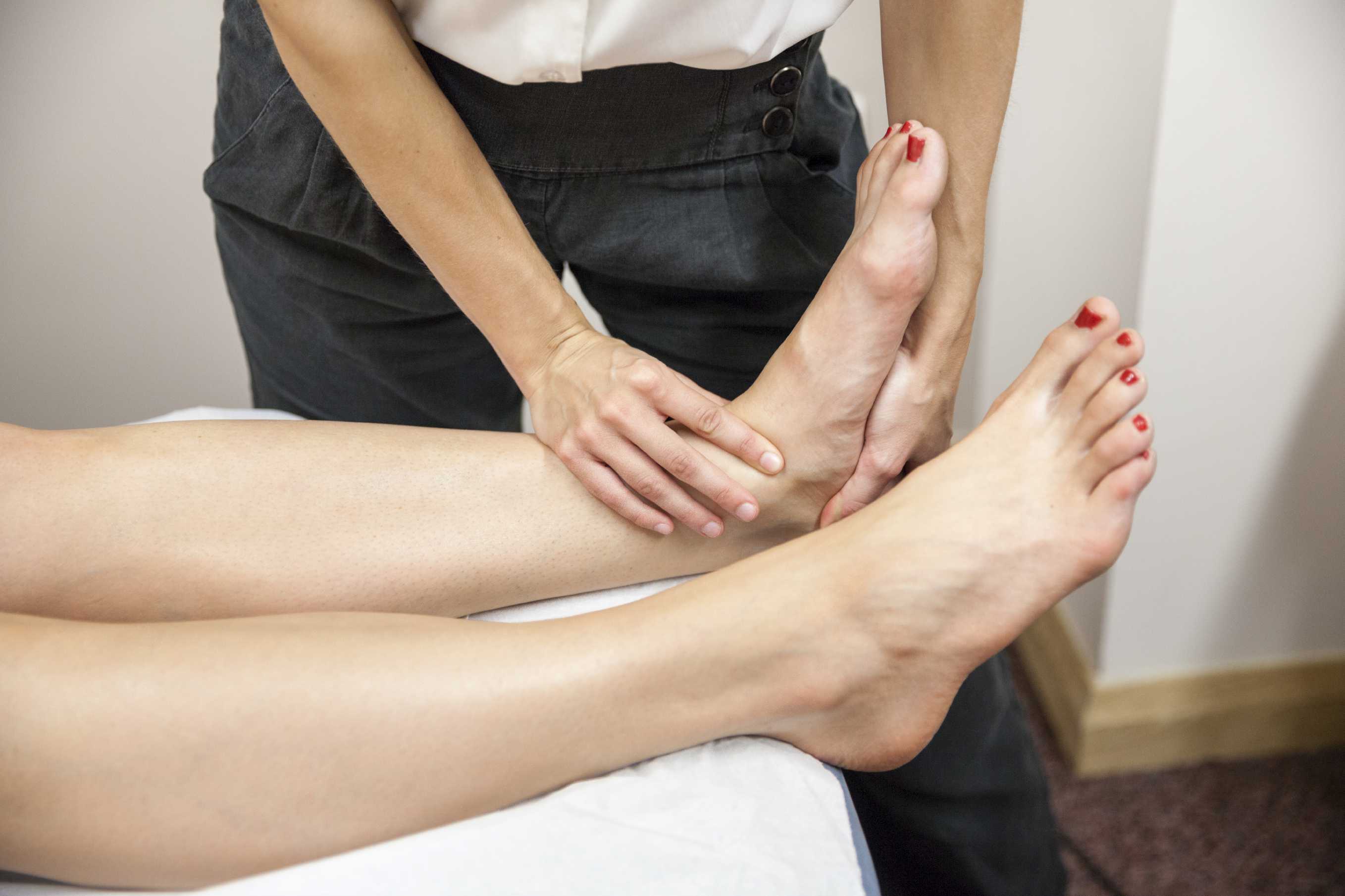
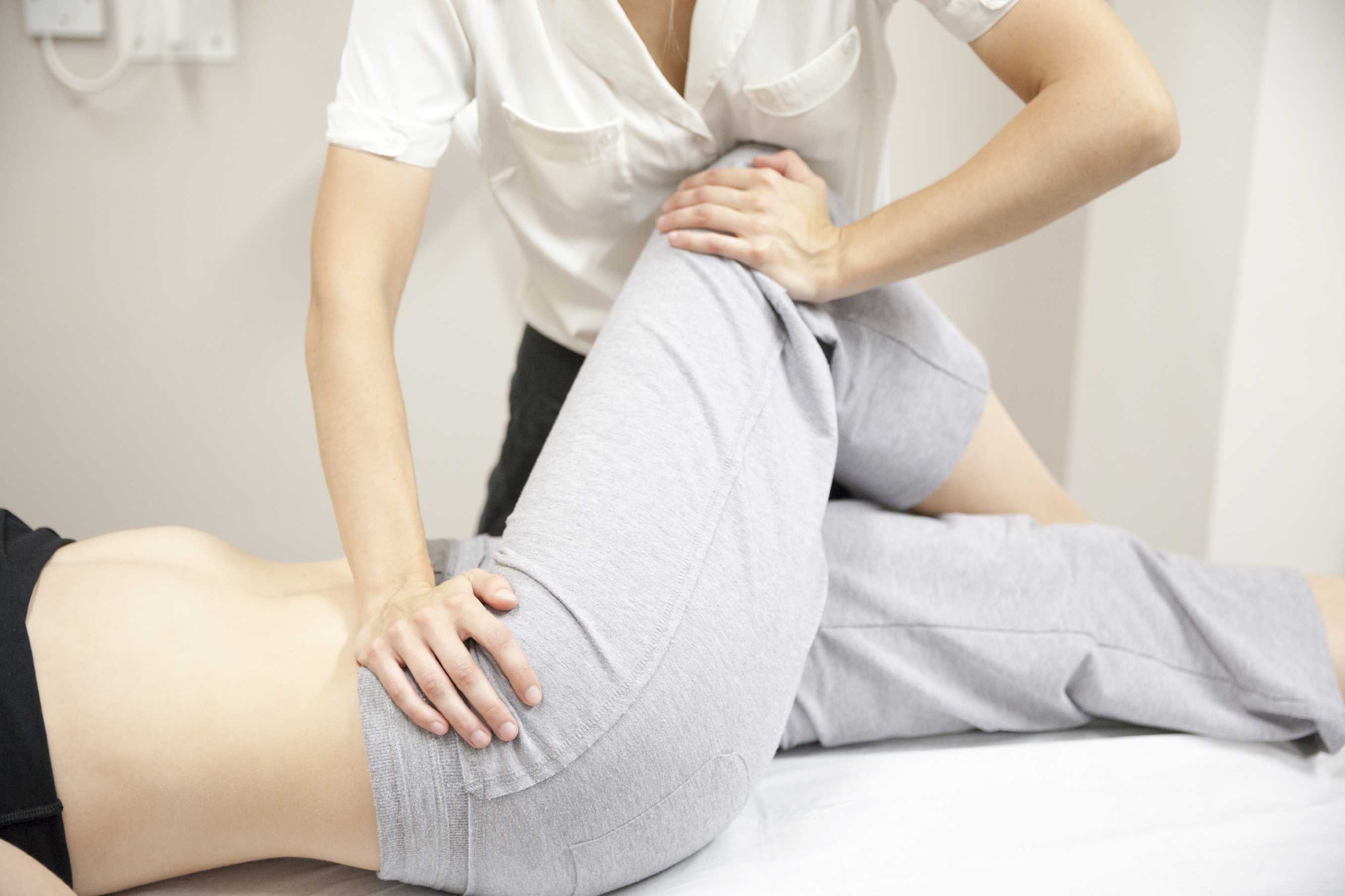
Physiotherapy management of spinal conditions:
No matter what your diagnosis or even if you are experiencing discomfort and you don’t know why, physiotherapy can help. We use a range of treatment techniques to help you to achieve your goals.
Treatment can include;
Diagnosed Conditions/Complaints – Peripheral
Injury- Tendon/Ligament/Muscle
These structures need to go through each stage of the healing process effectively to create new strong tissue that will withstand the forces that we place on our body in day to day life. This healing process can be broken down into three main phases and physiotherapy can help along the way:
The Inflammatory phase (acute and sub-acute);
This phase begins immediately after your injury with acute phase lasting from 0-7 days and the sub acute phase lasting from day 3 up to 3 weeks. This is a healthy response that clears any dead or damaged tissue and begins the repair process. Inflammation is necessary for healing but can become detrimental if excessive or prolonged. Your physiotherapist can assist in this acute stage by; advising on correct cryotherapy (use of ice) to minimise swelling, advise on the most appropriate ways to safely limit excessive movement to the injured tissue, provide appropriate exercises to maintain range of movement and aid circulation and if applicable electrotherapy may be used to expedite the healing process.
The Proliferative phase;
This stage overlaps with the inflammatory phase and is the process that restores tissue continuity via production of granulation tissue or scar tissue. Increased blood flow that is necessary to support the newly healed tissue is seen at the injury through a process called angiogenesis. At this stage it is important to start gentle strengthening exercises within a pain free range, this hastens optimal alignment of collagen fibres and promotes improved tissue mobility. Your physiotherapist can guide and monitor this process as it is vital that exercises are set at an appropriate level for you.
The Consolidation phase;
The initial repair phase produces what we call type III collagen which is relatively weak and has random orientation. As the healing process progresses the collagen becomes more aligned with local stresses and collagen III is gradually replaced with type I collagen that has greater tensile strength. This final stage will continue for months and often up to a year following the initial injury. Physical stress in the form of exercise is necessary to improve the strength in the repairing tissue. However it is suggested that this repaired structure will never match that of the pre-injury strength. At this stage you will be considering returning to sport or competition and rehabilitation to build on strength, flexibility, speed, endurance and balance is key to prevent re-injury.
Repetitive Strain Injury and Overuse
This encompasses a wide range of conditions that all cause pain and impairment and all arise through specific action or sustained postures required of them through work or sport. It is important to recognise that the work or sport is not the causative factor but there are often underlying variables that initiate the impairment which is further exacerbated by repetitive actions.
Risk factors include; failure to take adequate rest or breaks, inadequate training or poor technique, pressure to perform or complete a task, repetitive harmful or poor postures such as sitting or driving for long hours etc.
Unfortunately it can be difficult to ‘catch’ these injuries early as damage occurs over time at relatively mild peak forces. This means that you may not feel pain until some way down the line.
Treatment for these conditions requires guidance from your physiotherapist to identify key causative factors and to try to eliminate them. You may be able to treat your pain or injury but if the initiating factor, such as biomechanics or alignment is not corrected, the injury may return. Treatment may include specific strengthening or stretching exercises, soft tissue release and manual therapy to target areas of imbalance. Advice on posture, ergonomics, technique, rest and repair will also be provided which you should integrate in to your day to day life.
Tendinopathy
A tendinopathy is a specific over use or repetitive strain injury directly related to our tendons. Tendons have a low metabolic rate (oxygen consumption) to withstand loads and tension for long periods without the risk of ischaemia or cell death. This is an essential requirement of a structure joining muscle to bone. Unfortunately this adaptation can mean that they are slow to heal being poor in blood and nerve supply. Failure to heal results in subsequent tendinopathy as the integrity of the tendon is disrupted.
Clinical signs;
Pain related to load or activity. Initially pain may be present at the start of an activity but then reduce during the activity but may reappear on rest or when cooling down. Pain may change from a sharp pain to a dull ache after several weeks. One initial reaction to injury or pain is to rest but studies have shown that prolonged rest or ‘unloading’ of the tendon can be as harmful to tendons as overuse. Our aim is to assist your recovery and promote the quality of the repair so that you can return to your sport. We use a combination of techniques and electrotherapy to aid tissue healing and guide you with specific exercises such as an eccentric strengthening program, proven to reduce pain and improve the structural integrity of the tendon.
Arthrosis/Degenerative Conditions
The majority of joints in our body are what we call synovial joints. The ends of the bone are covered in smooth hyaline cartilage and surrounded by synovial fluid that facilitates smooth movement and absorbs impact. Damage to the joint earlier in life may predispose to arthrosis further down the line and is termed secondary arthrosis. You may develop primary arthrosis without any injury but it can be genetic or as a result of high BMI, sedentary lifestyle, muscle weakness or other factors.
Arthrosis is a process whereby the articular cartilage becomes thin, fissured and may break off. This exposes the underlying bone to increased forces and can cause sclerosis and micro-fracture. The synovial fluid and joint capsule may become inflamed due to joint debris causing heat, pain and swelling. Other changes within the joint can occur which can include reduced joint space between the two bones, alterations to the shape of the joint and development of bony spurs on the joint margin which may catch during movement.
This is a degenerative process that cannot be cured but it can be effectively managed so that pain is kept to a minimum and you are able to enjoy life and the hobbies that you love to do. The key to managing any degenerative condition is to maintain the strength and movement of the joint through exercise. This can often be easier said than done when you are experiencing pain! Your physiotherapist can use a range of treatment techniques to help you to reduce your pain often exacerbated by secondary compensations such as tight muscles. Manual techniques and mobilisations can help to improve range of movement, reduce pain and improve circulation of synovial fluid. Once your pain is more manageable you will be encouraged to begin a combination of strengthening exercises and low impact cardiovascular exercise. Degenerative conditions require a multi-disciplinary approach to be managed effectively which may include not just your physiotherapist but your doctor, podiatrist, dietician and occupational therapist.
Nerve entrapment/injury
Our peripheral nerves travel from the spinal cord to a muscle or organ following a pathway that weaves through and around bone, tendon, ligament and other muscles.
Nerve entrapment injuries result from chronic injury such as compression from the surrounding structures to the nerve at some point along this pathway. Injury to a nerve may result in ischaemia or lack of oxygen to the nerve which results in damage to the outside of the nerve. The outside of the nerve is known as the myelin sheath which is important in transmission of nerve signals. In severe cases the nerve may experience degeneration of the axons (centre of the nerve) and fibrotic changes that may prevent recovery.
You may experience symptoms such as; pain, burning sensation, tingling, numbness, pins and needles, muscle weakness.
Tight muscles, poor posture, abnormal joint movement, scar tissue, spinal pathology may all be a cause of nerve entrapment. Physiotherapy can help relieve these symptoms and treat this condition by releasing any restrictions that may be compressing the nerve. Nerve gliding exercises can help to ensure the flexibility of the nerve and allow it to slide through structures without restriction.
Fracture and Repair
Depending upon your injury your consultant may have advised a surgical repair or a cast as the best course of treatment for your fracture. You may also have been given some strict advice in regards to how much weight you can put through your leg or how much you can move your limb. It is vital that you take the advice from your consultant as the fracture site and healing process can be very delicate and easily delayed if disrupted. However irrespective of how much you can move your limb there are still exercises that will be safe to carry out. These will help to maintain muscle strength, prevent atrophy and maintain range of movement and flexibility until your restrictions are over. Gradually returning to full function can take some time, our aim would be to support this return to fitness through careful monitoring and a progressive rehab program tailored to your individual needs and goals.